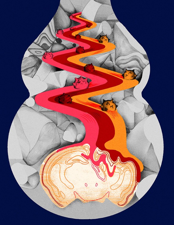
Maternal immune activation acts through interleukin-17a to promote ASD-like phenotypes in mice
Interleukin-17a (IL-17a) is a protein produced by activated T cells as part of the immune system’s response to infection. It promotes inflammation, and is associated with rheumatoid arthritis and psoriasis. Elevated IL-17a levels have been found in autistic children, and the IL17A gene is among many genes enriched in autistic patients. SCSB Investigators Gloria Choi and Jun Huh studied interleukin-17a (IL-17a) in mice, modeling maternal immune activation (MIA) during fetal development. Pregnant mice were subjected to immune system activation by injection of poly(I:C), which mimics viral infection.
Immune activation in the dams resulted in increased IL-17a mRNA expression in fetuses, abnormal cortical development, and communication and other behavioral changes in offspring. Direct application of IL-17a into fetal brain had similar effects, while all the effects could be rescued by blocking maternal IL-17a production. This study provides direct evidence of a role for MIA in the pathogenesis of ASD-like behavioral and cortical phenotypes in mice.
Learn more at the McGovern Institute or read the original research at Science (subscription content).


