Source: [Anne Trafton | MIT News, July 3, 2025]
MIT researchers found that low-quality visual input early in life may contribute to the development of key pathways in the brain’s visual system.
Incoming information from the retina is channeled into two pathways in the brain’s visual system: one that’s responsible for processing color and fine spatial detail, and another that’s involved in spatial localization and detecting high temporal frequencies. A new study from MIT provides an account for how these two pathways may be shaped by developmental factors.
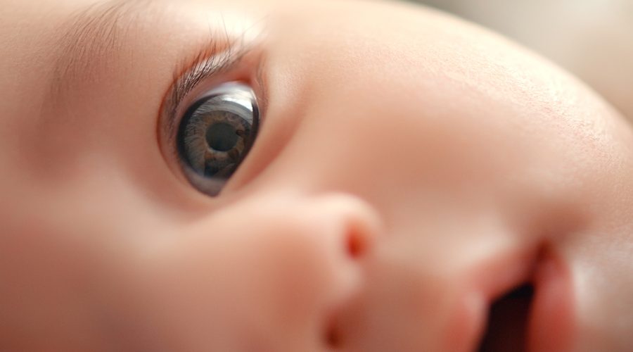
Newborns typically have poor visual acuity and poor color vision because their retinal cone cells are not well-developed at birth. This means that early in life, they are seeing blurry, color-reduced imagery. The MIT team proposes that such blurry, color-limited vision may result in some brain cells specializing in low spatial frequencies and low color tuning, corresponding to the so-called magnocellular system. Later, with improved vision, cells may tune to finer details and richer color, consistent with the other pathway, known as the parvocellular system.
To test their hypothesis, the researchers trained computational models of vision on a trajectory of input similar to what human babies receive early in life — low-quality images early on, followed by full-color, sharper images later. They found that these models developed processing units with receptive fields exhibiting some similarity to the division of magnocellular and parvocellular pathways in the human visual system. Vision models trained on only high-quality images did not develop such distinct characteristics.
“The findings potentially suggest a mechanistic account of the emergence of the parvo/magno distinction, which is one of the key organizing principles of the visual pathway in the mammalian brain,” says Pawan Sinha, an MIT professor of brain and cognitive sciences and the senior author of the study.
MIT postdocs Marin Vogelsang and Lukas Vogelsang are the lead authors of the study, which appears today in the journal Communications Biology. Sidney Diamond, an MIT research affiliate, and Gordon Pipa, a professor of neuroinformatics at the University of Osnabrueck, are also authors of the paper.
Sensory input
The idea that low-quality visual input might be beneficial for development grew out of studies of children who were born blind but later had their sight restored. An effort from Sinha’s laboratory, Project Prakash, has screened and treated thousands of children in India, where reversible forms of vision loss such as cataracts are relatively common. After their sight is restored, many of these children volunteer to participate in studies in which Sinha and his colleagues track their visual development.
In one of these studies, the researchers found that children who had cataracts removed exhibited a marked drop in object-recognition performance when the children were presented with black and white images, compared to colored ones. Those findings led the researchers to hypothesize that reduced color input characteristic of early typical development, far from being a hindrance, allows the brain to learn to recognize objects even in images that have impoverished or shifted colors.
“Denying access to rich color at the outset seems to be a powerful strategy to build in resilience to color changes and make the system more robust against color loss in images,” Sinha says.
In that study, the researchers also found that when computational models of vision were initially trained on grayscale images, followed by color images, their ability to recognize objects was more robust than that of models trained only on color images. Similarly, another study from the lab found that models performed better when they were trained first on blurry images, followed by sharper images.
To build on those findings, the MIT team wanted to explore what might be the consequences of both of those features — color and visual acuity — being limited at the outset of development. They hypothesized that these limitations might contribute to the development of the magnocellular and parvocellular pathways.
In addition to being highly attuned to color, cells in the parvocellular pathway have small receptive fields, meaning that they receive input from more compact clusters of retinal ganglion cells. This helps them to process fine detail. Cells in the magnocellular pathway pool information across larger areas, allowing them to process more global spatial information.
To test their hypothesis that developmental progressions could contribute to the magno and parvo cell selectivities, the researchers trained models on two different sets of images. One model was presented with a standard dataset of images that are used to train models to categorize objects. The other dataset was designed to roughly mimic the input that the human visual system receives from birth. This “biomimetic” data consists of low-resolution, grayscale images in the first half of the training, followed by high-resolution, colorful images in the second half.
After the models were trained, the researchers analyzed the models’ processing units — nodes within the network that bear some resemblance to the clusters of cells that process visual information in the brain. They found that the models trained on the biomimetic data developed a distinct subset of units that are jointly responsive to low-color and low-spatial-frequency inputs, similar to the magnocellular pathway. Additionally, these biomimetic models exhibited groups of more heterogenous parvocellular-like units tuned predominantly to higher spatial frequencies or richer color signals. Such distinction did not emerge in the models trained on full color, high-resolution images from the start.
“This provides some support for the idea that the ‘correlation’ we see in the biological system could be a consequence of the types of inputs that are available at the same time in normal development,” Lukas Vogelsang says.
Object recognition
The researchers also performed additional tests to reveal what strategies the differently trained models were using for object recognition tasks. In one, they asked the models to categorize images of objects where the shape and texture did not match — for example, an animal with the shape of cat but the texture of an elephant.
This is a technique several researchers in the field have employed to determine which image attributes a model is using to categorize objects: the overall shape or the fine-grained textures. The MIT team found that models trained on biomimetic input were markedly more likely to use an object’s shape to make those decisions, just as humans usually do. Moreover, when the researchers systematically removed the magnocellular-like units from the models, the models quickly lost their tendency to use shape to make categorizations.
In another set of experiments, the researchers trained the models on videos instead of images, which introduces a temporal dimension. In addition to low spatial resolution and color sensitivity, the magnocellular pathway responds to high temporal frequencies, allowing it to quickly detect changes in the position of an object. When models were trained on biomimetic video input, the units most tuned to high temporal frequencies were indeed the ones that also exhibited magnocellular-like properties in the spatial domain.
Overall, the results support the idea that low-quality sensory input early in life may contribute to the organization of sensory processing pathways of the brain, the researchers say. The findings do not rule out innate specification of the magno and parvo pathways, but provide a proof of principle that visual experience over the course of development could also play a role.
“The general theme that seems to be emerging is that the developmental progression that we go through is very carefully structured in order to give us certain kinds of perceptual proficiencies, and it may also have consequences in terms of the very organization of the brain,” Sinha says.
The research was funded by the National Institutes of Health, the Simons Center for the Social Brain, the Japan Society for the Promotion of Science, and the Yamada Science Foundation.