MRI scans reveal surprising similarities in activity patterns of infant and adult visual cortex.
Source: [Anne Trafton | MIT News Office, January 10, 2017]

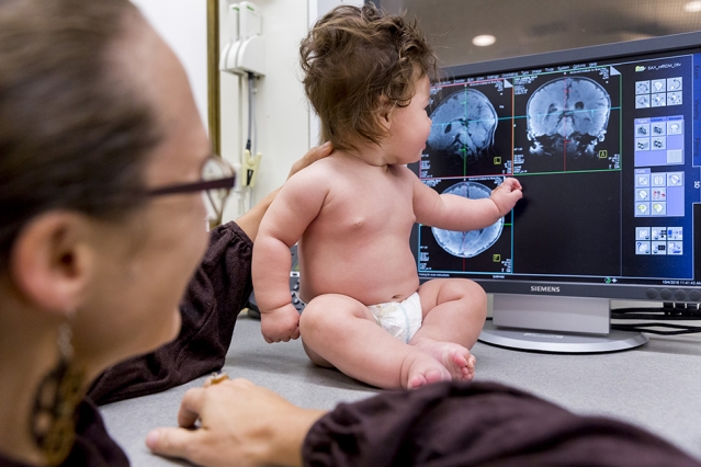
Neuroscientists at MIT have adapted their MRI scanner to make it easier to scan infants’ brains as the babies watch movies featuring different types of visual input. With the new technique, the team found that in some ways, the organization of infants’ brains is surprisingly similar to that of adults. Specifically, brain regions that respond to faces in adults do the same in babies, as do regions that respond to scenes.
Photo: Caitlin Cunningham
In adults, certain regions of the brain’s visual cortex respond preferentially to specific types of input, such as faces or objects — but how and when those preferences arise has long puzzled neuroscientists.
One way to help answer that question is to study the brains of very young infants and compare them to adult brains. However, scanning the brains of awake babies in an MRI machine has proven difficult.
Now, neuroscientists at MIT have overcome that obstacle, adapting their MRI scanner to make it easier to scan infants’ brains as the babies watch movies featuring different types of visual input. Using these data, the team found that in some ways, the organization of infants’ brains is surprisingly similar to that of adults. Specifically, brain regions that respond to faces in adults do the same in babies, as do regions that respond to scenes.
“It suggests that there’s a stronger biological predisposition than I would have guessed for specific cortical regions to end up with specific functions,” says Rebecca Saxe, a professor of brain and cognitive sciences and member of MIT’s McGovern Institute for Brain Research.
Saxe is the senior author of the study, which appears in the Jan. 10 issue of Nature Communications. The paper’s lead author is former MIT graduate student Ben Deen, who is now a postdoc at Rockefeller University.
MRI adaptations
Functional MRI (magnetic resonance imaging) is the go-to technique for studying brain function in adults. However, very few researchers have taken on the challenge of trying to scan babies’ brains, especially while they are awake.